What is an MRI?

Have you been diagnosed with a condition that requires you to undergo a procedure called MRI exam?
If you’re scheduled for a scan soon and would like to know more about this procedure, you’ve come to the right place.
MRI or Magnetic Resonance Imaging is a safe and non-invasive procedure for acquiring images inside the human body. Unlike traditional X-ray or CT scan, MRI does not use ionizing radiation.
If you want to know more about this state-of-the-art technology, read on.
WHAT IS AN MRI (MAGNETIC RESONANCE IMAGING)?

Magnetic resonance imaging (MRI), also known as nuclear magnetic resonance imaging, is a technique for creating detailed images of the human body.
The technique uses a very powerful magnet to align the nuclei of atoms inside the body and a variable magnetic field that causes the atoms to resonate. This phenomenon is called nuclear magnetic resonance.
The nuclei produce their own rotating magnetic fields that a scanner detects and uses to create an image.
An MRI scan can be used to examine different parts of the human body such as the:
- Brain and spinal cord
- Breasts
- Bones and joints
- Heart and blood vessels
- Internal organs like the liver, prostate gland, or womb
With the use of an MRI scanner, your conditions can be diagnosed, treatments can be planned and assessed if they are effective.
[Read: Defining Interventional Radiology: Meet Your Local IR Doctors]
What happens during an MRI scan?
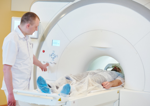
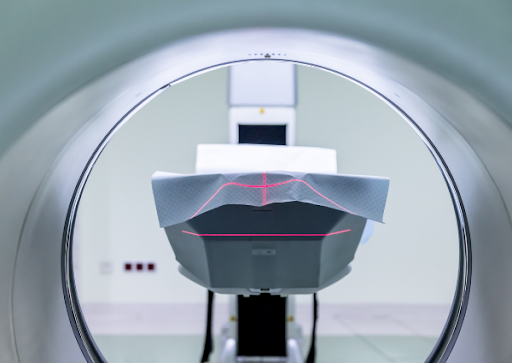
During an MRI scan, you lie down on a flat bed that is moved in the MRI machine. Depending on the part of your body that needs to be scanned, you are either positioned with your head or feet first.
The MRI machine is operated by a radiographer, who is trained in imaging and scans. They control the scanner using a computer located in a different room to keep it away from the magnetic fields and radio waves during an MRI procedure.
You can communicate with the radiographer through an intercom and they will be able to see you on a screen monitor throughout the MRI exam. You might hear loud tapping noises from the scanner during the procedure. This is the electric current in the scanner coils being turned on and off. You will be given earplugs or earphones to wear for your convenience.
It’s very important to keep as still as possible, with minimal movement, during the MRI scan. The procedure lasts 15 to 90 minutes, depending on the area of the body being scanned and also the number of detailed pictures being taken.
How does an MRI scan work?

The human body is made of water molecules, which consist of hydrogen and oxygen atoms. At the heart of each atom is a particle called a proton. Protons are like tiny magnets and are very sensitive to a magnetic field.
When you lie under the powerful scanner magnets, the protons in your body line up in the same direction similar to the way a compass needle aligns to a magnet. Short bursts of radio waves are then sent to certain locations on the body, knocking the protons out of alignment. When the radio waves are turned off, the protons realign. This sends out radio signals that are picked up by receivers.
These signals give information about the exact location of the protons in the body. They also give different signals about different tissues, because the protons realign at different speeds.
In the same way that millions of pixels can create complex pictures, the signals from the millions of protons in the body can provide detailed pictures of different parts of the body.
Uses of MRI
MRI is a non-invasive way for your doctor, through an MRI technician, to examine your organs, tissues, and skeletal system. It produces high-resolution images of the body that help diagnose different kinds of problems. These images are particularly important for a procedure such as a brain surgery where a functional MRI is used.
MRI of the brain and the spinal cord
MRI is often used to help diagnose:
- Aneurysms of cerebral vessels
- Multiple sclerosis
- Disorders of the inner ear and of the eye
- Stroke
- Brain injury from trauma
- Tumors
A special kind of MRI is the functional MRI of the brain or fMRI. It generates pictures of blood flow to certain points in the brain. It can be used to study brain anatomy and find out which parts of the brain are handling critical functions.
This helps identify important language as well as movement control areas of people considered for brain surgery. Functional MRI can also be used to evaluate damage from a head injury or conditions like Alzheimer’s disease.
MRI of the heart and blood vessels
MRI that focuses on the heart and blood vessels can check on the following:
- Thickness and movement of the walls of the heart
- Size and function of the chambers of the heart
- Structural problems in the aorta, like dissections or aneurysms
- Extent of damage caused by heart disease and heart attacks
- Inflammation or blockages in the blood vessels
MRI of other internal organs
MRI can check for abnormalities and tumors of many organs in the body, such as
- Kidneys
- Liver and bile ducts
- Pancreas
- Spleen
- Ovaries
- Uterus
- Prostate
MRI of bones and joints
MRI can also check:
- Disk abnormalities in the spine
- Joint abnormalities caused by repetitive or traumatic injuries like torn ligaments or cartilage
- Tumors of the bones and soft tissues
- Bone infections
[Read: Can Benign Tumors Become Cancerous? Interventional Radiologist Explains]
MRI of the breast
MRI, with mammography, can be used to detect breast cancer, especially in women who have dense breast tissue or who are at risk to have the condition.
MRI Safety
An MRI is a painless and safe procedure but you might find it uncomfortable if you have claustrophobia. In the MRI room, you will be advised to remove metal objects and metal or electronic devices. Also, if you have a metal implant fitted, such as a pacemaker or artificial joint, you might not be recommended to undergo an MRI scan.
As per the U.S. Food & Drug Administration, there are no known health hazards from temporary exposure to the MRI. Although it presents unique safety hazards for pregnant patients and those with implants, external devices, and accessory medical devices.
Learn more about MRI
MRI is a truly amazing and interesting technology with a vast number of medical uses. It is used in the diagnosis and treatment of injuries, diseases, and other medical conditions.
Generally, it is a safe and non-invasive procedure of getting detailed images of your body as long as it’s administered by a licensed MRI practitioner.
If you need consultation that requires the use of MRI in Prescott, Arizona, contact Vascular & Interventional Specialists of Prescott now!
Call us today at (928) 771-847.
Vascular & Interventional Specialists of Prescott was formed in 2010 by a group of subspecialty radiologists that perform numerous minimally-invasive, low-risk procedures using the tools of our trade for guidance—x-ray, ultrasound, CT scan, and MRI. The team’s goal is to educate patients and medical communities, while also providing safe and compassionate health care, with rapid recovery times and low risk of complications.
