MRI for Back Pain: What to Do Before, During, and After the Procedure
Having an MRI scan is probably not on the top of your list when it comes to how you want to spend your free time. But if you’re experiencing severe pain, your physician may require you to undergo this procedure.
In the case of back pain, most people would rather not undergo the procedure if they can avoid it altogether. However, when the doctor suspects that the underlying cause of your back pain is somewhat serious, your doctor might recommend an MRI. This is especially true if you have spinal pain along with leg and neck pain.
Continue reading if you want to know more about magnetic resonance imaging for back pain.
What is Magnetic Resonance Imaging?
Magnetic resonance imaging (MRI) is a safe and non-invasive diagnostic tool that makes use of radio waves and magnetic fields to produce detailed images of the body.
MRI was developed in the 80s and has revolutionized how severe back pain is diagnosed and treated. The technology is safe since the patient cannot feel the magnetic or radio waves. It does not use ionizing radiation that could potentially kill or damage cells.
MRI is For Diagnosis, Not Treatment
An MRI scan is typically considered to be the single best spinal imaging to help diagnose and treat back pain. It is considered one of the most vital medical devices today. Images produced by an MRI machine are important for the medical history of a patient.
MRI machines can also take pictures of soft tissues during a physical examination. Other symptoms and issues such as acute pain, herniated disk, bulging or ruptured disc, a previous spine injury, nerve root problems, back and leg pain, spinal stenosis (narrowing of spaces within the spine), and chest pain can be diagnosed better with the use of an MRI scanner.
When Is an MRI Scan Helpful for Back Pain
If the lumbar spine and the spinal cord are affected, there is definitely a need for MRI.
Health professionals may recommend an MRI if the back pain patient is exhibiting any of these symptoms:
- Inability to control urine or stools
- Cannot pass urine or stools
- Severe back pain that can’t be alleviated with home remedies and OTC medications
- Difficulty with balance and walking
- Fever
- History or any symptoms of cancer
- Recent serious fall or injury
- One leg feels weak or numb and is getting worse
[Related: Back Pain: Five Exercises That Can Lead to Long-Term Damage]
Before an MRI Scan
- You don’t need to do any time-consuming activity before you go to the hospital or radiology laboratory. A doctor or MRI technician will assess your medical history and provide any necessary procedure.
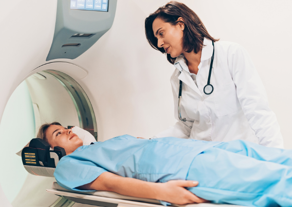
- The doctor will try to make you feel relaxed or calm. They will reassure you that you have nothing to worry about because an MRI scan is a safe procedure.
- For certain MRI exams, you may need to fast before the exam. If this is necessary, your doctor will notify you.
- You may be asked to take medication if you have had an MRI in the past and if you have had an allergic reaction to any of the contrast solutions. This will reduce the chance of you having another reaction.
- Talk to your doctor if you are claustrophobic or anxious about undergoing an MRI. The physician and radiologist will do the necessary adjustments to help you feel more relaxed.
- Your doctor may recommend sedation to calm you down. Let them know if you feel too drowsy.
- You will be asked to remove any metallic object that you wear, including hair clips, jewelry, watches, hearing aids, and dentures. Credit cards are also not allowed in the MRI room. Let your doctor know also if you have any metallic implants such as a pacemaker or a metal plate in your body.
During an MRI Scan
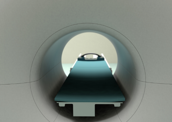
- The MRI machine is a long, narrow tube with both ends open. The tube opens and you can lie down on the movable table.
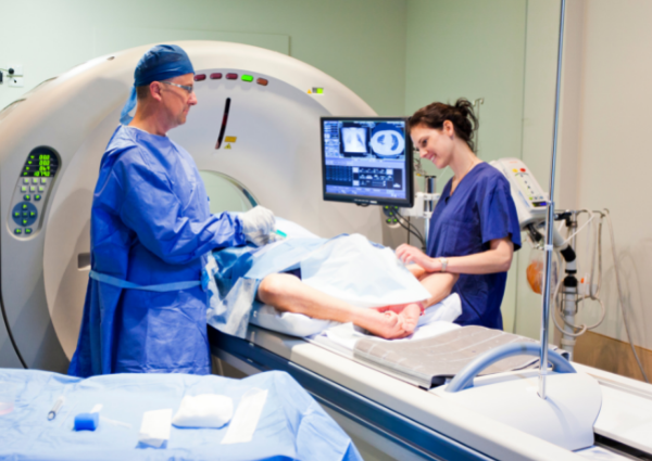
- From another room, an MRI technologist monitors your movements. The person can speak to you via microphone.
- You might be prescribed a drug to reduce anxiety and sleepiness if you are afraid of enclosed spaces (claustrophobia).
- The MRI machine creates strong magnetic fields around you and radio waves are directed toward your body. The procedure is safe and painless as there are no moving parts around and you can’t feel the magnetic field and radio waves.
- During the MRI scan, the magnet’s internal parts produce repetitive tapping, thumping, and other sounds when scanned. To help reduce the noise, you might be offered earplugs and/or music.
After an MRI Scan
- An MRI scan is done as an outpatient procedure. You won’t be required to stay overnight in a hospital for an MRI scan.
- You can immediately resume your normal activities after the scan.
- If you’ve taken sedatives, someone will need to bring you home. After taking a sedative, it is not safe to drive or operate heavy machinery for 24 hours.
- A radiologist will review your MRI scan and may also discuss it with other specialists.
- It is unlikely that you will receive the scan results immediately. The radiologist will send the report to the doctor who recommended the scan. They will then discuss the results with the patient.
- Unless they are urgently needed, it usually takes a week for results from an MRI scan to come through.
Where to Get MRI Scans for Back Pain in Prescott
An MRI scan is a non-invasive procedure that doctors use to help diagnose back pain, plan back surgery, or monitor progressive medical conditions like multiple sclerosis. The procedure is painless and there are very few risks or side effects.
MRI scans for back pain are outpatient procedures, so the person is usually free to leave the hospital or clinic after the examination.
If you need consultation that requires the use of MRI in Prescott, Arizona, contact Vascular & Interventional Specialists of Prescott now!
Call us today at (928) 771-847.
Vascular & Interventional Specialists of Prescott was formed in 2010 by a group of subspecialty radiologists that perform numerous minimally-invasive, low-risk procedures using the tools of our trade for guidance—x-ray, ultrasound, CT scan, and MRI. The team’s goal is to educate patients and medical communities, while also providing safe and compassionate health care, with rapid recovery times and low risk of complications.