What Are The Types of Peripheral Vascular Disease (PVD)?
Did you know that about 40 to 45 million adults in in the United States have been diagnosed with peripheral vascular disease (PVD)? Are you one of them too?
Peripheral vascular disease, also known as PVD, is a health issue that affects blood flow throughout our veins and arteries. It can present itself in many forms, and we’ll take a closer look about how this disease can manifest in a patient.
In this blog post, we’ll look at different types of Peripheral Vascular Disease (PVD), its symptoms and how you can recognize them if it ever strikes you or someone close.
What Is Peripheral Vascular Disease?
Peripheral Vascular Disease (PVD) is a circulatory disorder that involves the narrowing, blockage, or spasms of blood vessels outside of your heart and brain. It’s a slow and progressive circulation disorder that can affect any blood vessel in the body apart from those supplying the heart and brain.
Two Major Types of Peripheral Vascular Disease

According to Stanford Medicine, there are two major types of PVD – Occlusive PVD and Functional PVD. Both involve the most common peripheral vascular disease symptoms: blockage and narrowing of the blood vessels.
Let’s discuss these further below:
1. Occlusive Peripheral Vascular Disease
Under this type are medical conditions that cause blocking or narrowing of arteries that can lead to decreased blood flow.
Atherosclerosis
Due to a buildup of plaque on the artery walls, this medical disorder causes the arteries to narrow and stiffen. This condition can lead the plaques to burst and can progress into blood clots or a heart attack.
Symptoms:
- Chest pain or discomfort
- Pain in shoulders, back, neck, arms, and jaw
- Abdominal pain
- Shortness of breath
- Fatigue
- Confusion
Buerger’s Disease
Also known as thromboangiitis obliterans, this disease causes inflammation and swelling in the small and medium-sized blood vessel walls, typically in the legs and arms. When there is not enough blood flow, blood clots may form and may lead to death around the affected area, which is called gangrene.
Symptoms:
- Pain in the affected areas (hands and feet)
- Cold hands and feet
- Skin changes or appearance of small sores
Carotid Artery Disease
This condition occurs when the carotid arteries, the main blood vessels that carry oxygenated blood to the brain, become narrowed or blocked. Major risk factors of this disease include high blood pressure, high blood cholesterol, diabetes, family history of stroke, smoking, or trauma on the neck.
Symptoms:
- Sudden numbness or weakness in the face or limbs
- Trouble speaking or understanding speech
- Dizziness or loss of balance
- Sudden severe headache
- Brief loss of consciousness
Deep Vein Thrombosis (DVT)
This condition involves a blood clot forming in one or more of the deep veins in your body, usually in your legs. If a blood clot in your leg breaks loose, it can travel to your lungs and cause a pulmonary embolism, which can be life-threatening.
Symptoms:
- Leg pain or swelling
- Leg cramping
- Color changes on the leg
Lymphedema
This is a long-term condition where excess fluid collects in tissues causing swelling (edema). The condition usually affects the arms or legs, although it can affect other parts of the body.
Symptoms:
- Swelling in all or part of a limb or another part of the body
- Feeling of heaviness or tightness
- Restricted range of motion
- Aching or discomfort
- Hardening and thickening of the skin
2. Functional Peripheral Vascular Disease
The medical conditions under this category indicate that there is no current physical damage on the blood vessels’ structure. Instead, vessels widen and narrow in response to various factors like stress and temperature changes.
Chronic Venous Insufficiency (CVI)
CVI happens when the veins on your leg are damaged leading to too little blood flow back up to your heart, leading to blood pooling in the veins of your lower legs.
Symptoms:
- Swelling, aching, or tiredness in the legs
- Discoloration of the legs
- Hard, thick skin around the ankles
- Leg ulcers
Raynaud’s Disease
This is a condition that affects blood flow to the arteries of the fingers and toes, usually when exposed to cold temperatures or stress.
Symptoms:
- Cold fingers or toes
- Color changes in the skin in response to cold or stress
- Numb, prickly feeling or stinging pain upon warming or relief of stress
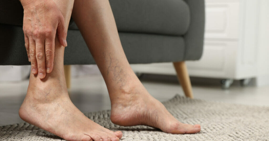
Varicose Veins:
These are enlarged, swollen, and twisting veins, often appearing blue or dark purple.
Symptoms:
- Aching leg
- Swollen feet and ankles
- Muscle cramp in legs
- Dry and itchy skin over the vein
- Skin discoloration
[Read More: Vein Specialist Explains: How to Get Rid of Varicose Veins Effectively]
Peripheral Vascular Disease vs. Peripheral Arterial Disease
Peripheral Vascular Disease (PVD) and Peripheral Arterial Disease (PAD) are often used interchangeably, but there is a subtle difference between the two.
- Peripheral Vascular Disease (PVD): This is a broader term that refers to diseases of any blood vessels located outside the heart and brain. PVD can affect both the arteries and veins. It includes conditions like peripheral artery disease, venous thrombosis (clots in the veins), and lymphatic diseases.
- Peripheral Arterial Disease (PAD): This is a subset of PVD that specifically involves problems with the arterial system, or the blood vessels that carry blood away from the heart to the body. PAD is typically caused by atherosclerosis, a buildup of plaque that narrows or blocks the arteries, reducing blood flow to the limbs.
[Read More: What Is the Difference Between PAD and PVD?]
What Causes Peripheral Vascular Disease?
Several risk factors have been associated with PVD. These include:
- Smoking: This is the strongest and most common risk factor for PVD. It increases the risk of developing PVD by several fold, and smokers tend to develop PVD earlier than non-smokers.
- Diabetes: People with diabetes are at a higher risk of developing PVD.
- Age: PVD mostly occurs in people over age 60.
- Prior coronary artery disease: Individuals who have previously suffered from coronary artery disease are more likely to develop PVD.
How Is Peripheral Vascular Disease Diagnosed?
Peripheral Vascular Disease (PVD) is usually diagnosed based on your medical and family history, a physical exam, and results from tests. Here are some common methods used to diagnose PVD:
- Ankle-Brachial Index (ABI): It compares the blood pressure in your ankle to the blood pressure in your arm.
- Angiogram: This test uses X-ray imaging to view your blood vessels. A special dye is injected into the blood vessels, making them visible on the X-ray.
- Segmental Doppler Pressure Testing: This method checks different parts of your legs for narrowed or blocked arteries using blood pressure cuffs placed at thigh, calf, and ankle levels.
Have Your Arteries and Veins Checked By Our Specialists

PVD can manifest in various medical conditions, and it is essential to get treated by our vascular specialists at VISP. We commit to providing you with safe and minimally invasive procedures to cure or prevent peripheral vascular disease and peripheral artery disease.
Contact us today to set an appointment!
Vascular & Interventional Specialists of Prescott was formed in 2010 by a group of subspecialty radiologists that perform numerous minimally-invasive, low-risk procedures using the tools of our trade for guidance—x-ray, ultrasound, CT scan, and MRI. The team’s goal is to educate patients and medical communities, while also providing safe and compassionate health care, with rapid recovery times and low risk of complications.