What Happens During a Pelvic Congestion Syndrome Ultrasound?
A lot of women worldwide are suffering from significant and debilitating pelvic pain that could be due to a condition known as pelvic congestion syndrome.
In fact, approximately one-third of all women suffer from chronic pelvic pain at least at one point during their lifetime. Pelvic congestion syndrome (PVC) is caused by varicose veins deep within the pelvic region.
PVC could go undiagnosed and remain essentially untreated if not paid careful attention. If you are among those who experience chronic pelvic pain, read on to know more about it.
What is Pelvic Congestion Syndrome?

Chronic pelvic pain can happen for many reasons including but not limited to an ovarian cyst, irritable bowel syndrome, musculoskeletal problems, painful bladder, other gynecological problems as well as psychological factors.
Also known as pelvic venous insufficiency, pelvic congestion syndrome is a vascular disorder that affects the pelvic veins and is similar to varicose veins.
The condition occurs when there is no proper drainage of blood in the region. It is most common in women who have given birth more than once and causes clinical symptoms such as painful, swollen, and bulging pelvic and ovarian veins, also known as pelvic varicose veins.
The symptoms have to be chronic, meaning lasting for 6 months or more. Vascular tests and risk factors have to be assessed before a medical doctor determines that the disorder is related to the circulatory system. Risk factors include age, obesity, previous pregnancy, and family history.
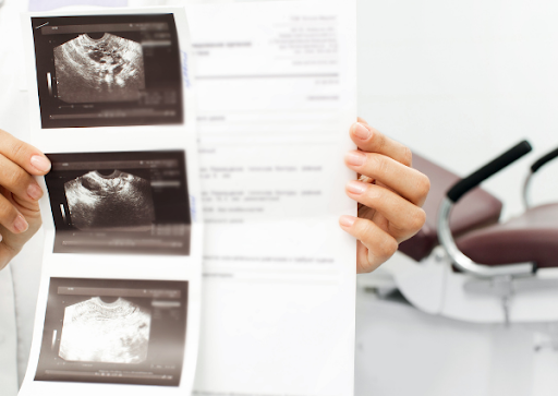
Major symptoms are usually assessed with a pelvic ultrasound and a vascular technician. A complete evaluation of a gynecologist has to be completed to rule out any underlying gynecological causes of the symptoms.
[Related: Varicose Veins: Pain, Swelling & Managing Your Symptoms]
CAUSES
Normal veins are those that have blood flowing from the pelvis into the heart in the ovarian vein. The vein has valves that prevent blood from flowing backward.
When the ovarian valve is damaged or weak, blood cannot flow efficiently from the vein into the heart. This causes blood to reflux and pool in the vein, leading to congestion and swelling.
The result? Chronic pain, heaviness, and pelvic varicose veins.
Numerous events can cause damage to the pelvic vessels, including:
- Weight gain
- Hormonal fluctuations
- Multiple pregnancies
Injured pelvic veins may cause swelling in surrounding organs, such as the bladder and intestines. Most women affected by PCS are between the ages of 20 and 45, with multiple pregnancies.
During pregnancy, hormones fluctuate, pelvic anatomy changes, and the volume of blood and estrogen increases. These conditions can weaken the walls of the blood vessels.
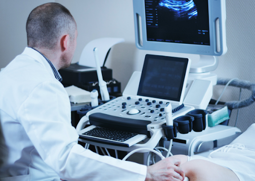
What is an ultrasound of the pelvis?
Ultrasound imaging (sonography) is a non-invasive medical test that helps physicians diagnose as well as treat medical conditions. It is safe and painless. It generates images of the body with the use of sound waves. It makes use of a small probe called a transducer and gel placed directly on the skin.
High-frequency waves travel from the probe, through the gel on the skin, and then into the body. The probe collects the sound waves that bounce back. A computer uses those sound signals to produce images.
Sonography does not involve radiation such as X-rays. Because ultrasound captures images in real-time, it can show the movement and structure of organs as well as blood flow.
Ultrasound imaging of the pelvis uses sound waves to generate images of the structures and organs in the pelvis and the lower abdomen. There are three kinds of pelvic ultrasound – abdominal, vaginal (for women), and rectal (for men). These exams are frequently non-invasive and are used to evaluate the reproductive and urinary systems.
During pelvic sonography, the practitioner will examine if there’s gonadal vein reflux (ovarian vein for women, and testicular vein for men).
Aside from a dilated gonadal vein, they will also search for anatomical and hemodynamic criteria of left renal vein compression, examine the external and internal iliac veins, and watch out for any leaks.
In general, the procedure is safe and does not use ionizing radiation. This procedure requires little to no preparation.
You might be asked to hydrate or drink water before the examination to fill your bladder and improve your blood flow. Leave your jewelry at home and wear loose, comfortable clothing. You might also be asked to wear a gown.
A Doppler ultrasound exam could be part of a pelvic ultrasound examination. It is a special ultrasound procedure that detects the movement of material in the body. It lets the doctor see the movement in the arteries as well as blood flow through the blood vessels such as the uterine veins, ovarian vein, and gonadal vein.
What are some common uses of pelvic congestion syndrome ultrasound?
In women, a pelvic ultrasound is used to diagnose the:
- Uterus
- Ovaries
- Bladder
- Fallopian tubes
- Cervix
It can be also used to monitor the health and development of a fetus or an embryo during pregnancy. It may be also used to diagnose:
- Pelvic pain
- Infertility
- Other menstrual problems
- Abnormal vaginal bleeding
GET PCS TREATMENT FROM A TRUSTED VEIN DOCTOR
Additional diagnostic tests may be recommended depending on the severity and nature of your pain. Once pelvic congestion syndrome is confirmed, ovarian vein embolization may be performed.
Ovarian Vein Embolization is a minimally invasive procedure that can relieve pain and other symptoms It involves placing a small catheter into the enlarged pelvic veins through a tiny incision using x-ray guidance.
[For detailed treatment information, read: Vein Specialist Explains Pelvic Congestion Syndrome Treatment]
Vascular & Interventional Specialists of Prescott (VISP) has been a part of the Prescott medical community and serving patients since 2010. We are a group of subspecialty radiologists that perform numerous minimally-invasive, low-risk procedures using the tools of our trade for guidance—x-ray, ultrasound, CT scan, and MRI.
If you’re experiencing any symptoms of pelvic congestion syndrome, don’t hesitate to visit our clinic for an in-depth health consultation.
To book a doctor’s appointment for pelvic congestion syndrome, call us at 928.771.8477.
Vascular & Interventional Specialists of Prescott was formed in 2010 by a group of subspecialty radiologists that perform numerous minimally-invasive, low-risk procedures using the tools of our trade for guidance—x-ray, ultrasound, CT scan, and MRI. The team’s goal is to educate patients and medical communities, while also providing safe and compassionate health care, with rapid recovery times and low risk of complications.